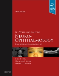
Liu, Volpe, Galetta's Neuro-Ophthalmology: Diagnosis
and Management, 2019, 3rd
edition
Editors: Liu GT, Volpe NJ,
Galetta SL
Publisher: Elsevier
(www.elsevier.com
for more information)


Review of the first edition, by
Anderson D. New England
Journal of Medicine 2001;344:1872-1873. 
Review of the second edition, by Jay W. JAMA 2011;306:439-440. 
Review of the third edition, by Golnik KC. J Neuro-ophthalmol 2018;38(4):e22. 
This book is designed for the students of neuro-ophthalmology:
the practitioners, ophthalmology and neurology residents, and
medical students dealing with patients with disturbances of the
afferent visual pathways and efferent ocular motor systems.
Our hope was to offer a book that
could serve both as a guide for diagnosing and managing patients
with neuro-ophthalmic problems and a one-volume textbook containing
in-depth discussions of neuro-ophthalmic topics and disorders. Our aim was to create a highly organized
and uniform textbook, extensively illustrated and referenced,
that would bridge the gap between neuro-ophthalmic handbooks containing
tables, outlines, and flow-diagrams and neuro-ophthalmic encyclopedias. The third edition, enhanced by contributions from former Penn Neuro-Ophthalmology fellows, has over 1,000 illustrations and almost 200 videos.
How the book is organized (see below
for table of contents)
The book is divided into four
sections: history and examination, afferent disorders, efferent
disorders, and headache. This initial grouping of the last three
sections is symptom-based, and within each of those sections,
the chapters then are divided neuroanatomically . Each chapter
has roughly the same internal organization and starts with an
introduction to the structures or disorders discussed, followed
by a review of their neuroanatomy, symptoms, and signs. This is
followed by a detailed discussion of the presentation, pathophysiology,
diagnosis, neuroimaging and diagnostic studies, and management
of the diseases which affect that structure.
Within the chapters, the liberal
use of headings such as "neuro-ophthalmic manifestations"
points the reader to exactly what he or she may want to know about
a particular topic. The copious use of figures and tables aids
the discussions, and the generous references direct those desiring
to pursue a topic in greater depth. Repetition between chapters,
sometimes unavoidable for the sake of completeness, was kept to
a minimum by cross-referencing topics, figures, and tables. Because
this is a book for clinicians, reviews of neuroanatomy and
neurophysiology
in most instances are based upon clinical and pathological observations
in humans without extensive discussion of experimental literature
involving nonhuman primates and other animals.
The first two chapters on history
and examination were designed for students and residents unfamiliar
with eliciting symptoms and signs in neuro-ophthalmic patients.
The examination chapter also includes reviews of the neurological
examination and the bedside neuro-ophthalmic evaluation of comatose
patients. Often based on the history alone, the problem can be
localized neuroanatomically and its etiology determined. The
examination
can then be used to support a suspected diagnosis and eliminate
other causes.
The next section on afferent disorders
discusses the diseases associated with structures responsible
for transmitting and processing visual information coming from
the eye. Here disorders of the retina, optic nerve, chiasm, and
retrochiasmal structures, as well as disc swelling, higher cortical
disorders, transient and functional visual loss, and hallucination
and illusions are discussed. Readers unfamiliar with these subjects
or topical localization of visual field defects might first review
Chapter 3, which contains an overview of afferent visual disorders
and visual field testing.
The section on efferent disorders
reviews conditions involving motor neuro-ophthalmic structures.
Thus pupillary, eyelid, and facial nerve abnormalities, then eye
movement and orbital disorders are discussed here. Eye movement
disorders are covered in four chapters: Chapter 15 reviews causes
of dysconjugate gaze including third, fourth, and sixth nerve
palsies; Chapter 16 discusses abnormalities of conjugate gaze
such as defective saccades and pursuit; Chapter 17 outlines conditions
where the eyes move excessively as in nystagmus and nystagmoid
movements; and Chapter 18 discusses eye movement and other
abnormalities
in orbital disorders.
Chapter 19: headache, faceache,
and disorders of facial sensation discusses topics such as migraine,
cluster headache, trigeminal neuralgia, and trigeminal neuropathy.
Table of Contents
i) Introduction
I. History and Examination
1. Neuro-ophthalmic History
2. Neuro-ophthalmic Examination
II. Visual Loss and Other Disorders
of the Afferent Visual Pathway
3. Visual Loss: Overview, Visual Field Testing, and Topical Diagnosis
4. Visual Loss: Retinal Disorders of Neuro-ophthalmic Interest
5. Visual Loss: Optic Neuropathies
6. Optic Disc Swelling: Papilledema and Other Causes
7. Visual Loss: Chiasmal Disorders
8. Visual Loss: Retrochiasmal Disorders
9. Disorders of Higher Cortical Visual Function
10. Transient Visual Loss or Blurring
11. Functional (Nonorganic) Visual Loss
12. Visual Hallucinations and Illusions
III. Efferent Neuro-Ophthalmic
Disorders
13. Pupillary Disorders
14. Eyelid and Facial Nerve Disorders
15. Eye Movement Disorders: Third, Fourth, and Sixth Nerve Palsies
and Other
Causes of Diplopia and Ocular Misalignment
16. Eye Movement Disorders: Gaze Abnormalities
17. Eye Movement Disorders: Nystagmus and Nystagmoid Eye Movements
18. Orbital Disease in Neuro-ophthalmology
IV. Other Topics
19. Headache, Faceache, and Disorders of Facial Sensation


